Electroencephalography
Contents
Electroencephalography#
Support for Electroencephalography (EEG) was developed as a BIDS Extension Proposal. Please see Citing BIDS on how to appropriately credit this extension when referring to it in the context of the academic literature.
Several example EEG datasets have been formatted using this specification and can be used for practical guidance when curating a new dataset.
EEG recording data#
The EEG community uses a variety of formats for storing raw data, and there is no single standard that all researchers agree on. For BIDS, EEG data MUST be stored in one of the following formats:
Format |
Extension(s) |
Description |
|---|---|---|
|
Each recording consists of a single |
|
|
Each recording consists of a |
|
|
The format used by the MATLAB toolbox EEGLAB. Each recording consists of a |
|
|
Each recording consists of a single |
It is RECOMMENDED to use the European data format, or the BrainVision data
format. It is furthermore discouraged to use the other accepted formats over
these RECOMMENDED formats, particularly because there are conversion scripts
available in most commonly used programming languages to convert data into the
RECOMMENDED formats. The data in their original format, if different from the
supported formats, can be stored in the /sourcedata directory.
The original data format is especially valuable in case conversion elicits the
loss of crucial metadata specific to manufacturers and specific EEG systems. We
also encourage users to provide additional meta information extracted from the
manufacturer specific data files in the sidecar JSON file. Other relevant files
MAY be included alongside the original EEG data in /sourcedata.
Note the RecordingType, which depends on whether the data stream on disk is interrupted or not. Continuous data is by definition 1 segment without interruption. Epoched data consists of multiple segments that all have the same length (for example, corresponding to trials) and that have gaps in between. Discontinuous data consists of multiple segments of different length, for example due to a pause in the acquisition.
Note that for proper documentation of EEG recording metadata it is important to understand the difference between electrode and channel: An EEG electrode is attached to the skin, whereas a channel is the combination of the analog differential amplifier and analog-to-digital converter that result in a potential (voltage) difference that is stored in the EEG dataset. We employ the following short definitions:
Electrode = A single point of contact between the acquisition system and the recording site (for example, scalp, neural tissue, …). Multiple electrodes can be organized as caps (for EEG), arrays, grids, leads, strips, probes, shafts, and so on.
Channel = A single analog-to-digital converter in the recording system that regularly samples the value of a transducer, which results in the signal being represented as a time series in the digitized data. This can be connected to two electrodes (to measure the potential difference between them), a magnetic field or magnetic gradient sensor, temperature sensor, accelerometer, and so on.
Although the reference and ground electrodes are often referred to as channels, they are in most common EEG systems not recorded by themselves. Therefore they are not represented as channels in the data. The type of referencing for all channels and optionally the location of the reference electrode and the location of the ground electrode MAY be specified.
Sidecar JSON (*_eeg.json)#
Generic fields MUST be present:
SHOULD be present: For consistency between studies and institutions, we encourage users to extract the values of these fields from the actual raw data. Whenever possible, please avoid using ad hoc wording.
Specific EEG fields MUST be present:
SHOULD be present:
Example:
{
  "TaskName":"Seeing stuff",
  "TaskDescription":"Subjects see various images for which phase, amplitude spectrum, and color vary continuously",
  "Instructions":"Your task is to detect images when they appear for the 2nd time, only then press the response button with your right/left hand (counterbalanced across subjects)",
  "InstitutionName":"The world best university, 10 Beachfront Avenue, Papeete",
  "SamplingFrequency":2400,
  "Manufacturer":"Brain Products",
  "ManufacturersModelName":"BrainAmp DC",
  "CapManufacturer":"EasyCap",
  "CapManufacturersModelName":"M1-ext",
  "EEGChannelCount":87,
  "EOGChannelCount":2,
  "ECGChannelCount":1,
  "EMGChannelCount":0,
  "MiscChannelCount":0,
  "TriggerChannelCount":1,
  "PowerLineFrequency":50,
  "EEGPlacementScheme":"10 percent system",
  "EEGReference":"single electrode placed on FCz",
  "EEGGround":"placed on AFz",
  "SoftwareFilters":{
    "Anti-aliasing filter":{
      "half-amplitude cutoff (Hz)": 500,
      "Roll-off": "6dB/Octave"
    }
  },
  "HardwareFilters":{
    "ADC's decimation filter (hardware bandwidth limit)":{
      "-3dB cutoff point (Hz)":480,
      "Filter order sinc response":5
    }
  },
  "RecordingDuration":600,
  "RecordingType":"continuous"
}
Note that the date and time information SHOULD be stored in the Study key file
(scans.tsv).
Date time information MUST be expressed as indicated in Units
Channels description (*_channels.tsv)#
This file is RECOMMENDED as it provides easily searchable information across BIDS datasets. For example for general curation, response to queries, or for batch analysis. To avoid confusion, the channels SHOULD be listed in the order they appear in the EEG data file. Any number of additional columns MAY be added to provide additional information about the channels.
Note that electrode positions SHOULD NOT be added to this file, but to
*_electrodes.tsv.
Furthermore, the entries in *_electrodes.tsv and *_channels.tsv do not have to match exactly,
as for example in the case of recording a single EOG channel from a bipolar referencing scheme
of two electrodes, or a data channel originating from an auxiliary, non-electrode device.
That is, in most cases *_electrodes.tsv will have more entries than *_channels.tsv.
See the examples for *_channels.tsv below, and for *_electrodes.tsv in
“Electrodes description”.
The columns of the channels description table stored in *_channels.tsv are:
MUST be present in this specific order:
SHOULD be present:
Restricted keyword list for field type in alphabetic order (shared with the
MEG and iEEG modality; however, only the types that are common in EEG data are listed here).
Note that upper-case is REQUIRED:
Keyword |
Description |
|---|---|
AUDIO |
Audio signal |
EEG |
Electroencephalogram channel |
EOG |
Generic electrooculogram (eye), different from HEOG and VEOG |
ECG |
Electrocardiogram (heart) |
EMG |
Electromyogram (muscle) |
EYEGAZE |
Eye tracker gaze |
GSR |
Galvanic skin response |
HEOG |
Horizontal EOG (eye) |
MISC |
Miscellaneous |
PPG |
Photoplethysmography |
PUPIL |
Eye tracker pupil diameter |
REF |
Reference channel |
RESP |
Respiration |
SYSCLOCK |
System time showing elapsed time since trial started |
TEMP |
Temperature |
TRIG |
System triggers |
VEOG |
Vertical EOG (eye) |
Example of free-form text for field description
n/a, stimulus, response, skin conductance, battery status
Example channels.tsv#
See also the corresponding electrodes.tsv example.
name     type  units  description                     reference     status  status_description
VEOG     VEOG  uV     left eye                        VEOG-, VEOG+  good    n/a
FDI      EMG   uV     left first dorsal interosseous  FDI-, FDI+    good    n/a
Cz       EEG   uV     n/a                             REF           bad     high frequency noise
UADC001  MISC  n/a    envelope of audio signal        n/a           good    n/a
Electrodes description (*_electrodes.tsv)#
File that gives the location of EEG electrodes. Note that coordinates are
expected in cartesian coordinates according to the EEGCoordinateSystem and
EEGCoordinateUnits fields in *_coordsystem.json. If an
*_electrodes.tsv file is specified, a *_coordsystem.json
file MUST be specified as well. The order of the required columns in the
*_electrodes.tsv file MUST be as listed below.
MUST be present in this specific order:
SHOULD be present:
Example electrodes.tsv#
See also the corresponding electrodes.tsv example.
name   x        y	       z        type     material
VEOG+  n/a      n/a      n/a      cup      Ag/AgCl
VEOG-  n/a      n/a      n/a      cup      Ag/AgCl
FDI+   n/a      n/a      n/a      cup      Ag/AgCl
FDI-   n/a      n/a      n/a      cup      Ag/AgCl
GND    -0.0707  0.0000   -0.0707  clip-on  Ag/AgCl
Cz     0.0000   0.0714   0.0699   cup      Ag/AgCl
REF    -0.0742  -0.0200  -0.0100  cup      Ag/AgCl
The acq-<label> key/value pair can be used to indicate acquisition of the same data. For
example, this could be the recording of electrode positions with a different
electrode position recording device, or repeated digitization before and after
the recording.
Coordinate System JSON (*_coordsystem.json)#
A *_coordsystem.json file is used to specify the fiducials, the location of
anatomical landmarks, and the coordinate system and units in which the position
of electrodes and landmarks is expressed. The *_coordsystem.json is
REQUIRED if the optional *_electrodes.tsv is specified. If a corresponding
anatomical MRI is available, the locations of landmarks and fiducials according
to that scan should also be stored in the *_T1w.json
file which goes alongside the MRI data.
For disambiguation, we employ the following definitions for fiducials and anatomical landmarks respectively:
Fiducials are objects with a well defined location used to facilitate the localization of electrodes and co-registration with other geometric data such as the participant’s own T1 weighted magnetic resonance head image, a T1 weighted template head image, or a spherical head model. Commonly used fiducials are vitamin-E pills, which show clearly in an MRI, or reflective spheres that are localized with an infrared optical tracking system.
Anatomical landmarks are locations on a research subject such as the nasion, which is the intersection of the frontal bone and two nasal bones of the human skull.
Fiducials are typically used in conjunction with anatomical landmarks. An example would be the placement of vitamin-E pills on top of anatomical landmarks, or the placement of LEDs on the nasion and preauricular points to triangulate the position of other LED-lit electrodes on a research subject’s head.
For more information on the definition of anatomical landmarks, please visit: https://www.fieldtriptoolbox.org/faq/how_are_the_lpa_and_rpa_points_defined
For more information on coordinate systems for coregistration, please visit: https://www.fieldtriptoolbox.org/faq/how_are_the_different_head_and_mri_coordinate_systems_defined
General fields:
Fields relating to the EEG electrode positions:
Fields relating to the position of fiducials measured during an EEG session/run:
Fields relating to the position of anatomical landmark measured during an EEG session/run:
If the position of anatomical landmarks is measured using the same system or
device used to measure electrode positions, and if thereby the anatomical
landmarks are expressed in the same coordinates, the coordinates of the
anatomical landmarks can be specified in electrodes.tsv. The same applies to
the coordinates of the fiducials.
Anatomical landmarks or fiducials measured on an anatomical MRI that match the
landmarks or fiducials during an EEG session/run, must be stored separately in
the corresponding *_T1w.json or *_T2w.json file and should be expressed in
voxels (starting from [0, 0, 0]).
Example:
{
  "IntendedFor":"/sub-01/ses-01/anat/sub-01_T1w.nii",
  "EEGCoordinateSystem":"Other",
  "EEGCoordinateUnits":"mm",
  "EEGCoordinateSystemDescription":"RAS orientation: Origin halfway between LPA and RPA, positive x-axis towards RPA, positive y-axis orthogonal to x-axis through Nasion,  z-axis orthogonal to xy-plane, pointing in superior direction.",
  "FiducialsDescription":"Electrodes and fiducials were digitized with Polhemus, fiducials were recorded as the centre of vitamin E capsules sticked on the left/right pre-auricular and on the nasion, these are also visible on the T1w MRI"
}
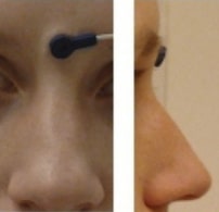
Landmark photos (*_photo.jpg)#
Photos of the anatomical landmarks and/or fiducials.
Photos of the anatomical landmarks and/or fiducials are OPTIONAL. Please note that the photos may need to be cropped or blurred to conceal identifying features prior to sharing, depending on the terms of the consent given by the participant.
The acq-<label> key/value pair can be used to indicate acquisition of different photos of
the same face (or other body part in different angles to show, for example, the
location of the nasion (NAS) as opposed to the right periauricular point (RPA).
Example:
Picture of a NAS fiducial placed between the eyebrows, rather than at the
actual anatomical nasion: sub-0001_ses-001_acq-NAS_photo.jpg